
Your prenatal care at the American Hospital of Paris includes:
- Prenatal visits with your midwife or obstetrician-gynecologist, until your due date
- Early prenatal interview with a midwife from our team
- Anesthesia consultation at the end of your pregnancy. (Even if you have chosen to forgo anesthesia, in some cases it can become necessary during childbirth. This consultation ensures the anesthesia team has your complete medical file if necessary, which guarantees maximum safety for you.)
- At least three obstetric screening ultrasounds performed by our Fetal Medicine Unit
The arrival of a baby requires that you be prepared, and we are here to provide the personalized support you need. Because we talk about you and your baby during our staff meetings, which are attended by the fetal medicine, obstetric and pediatric experts involved in the birth, we know you and your unborn child and prepare for the delivery based on your respective needs.
The American Hospital of Paris provides unparalleled personalized support to you throughout this journey. Our team takes care to inform you about your state of health, and that of your baby, so you can participate in decisions about your prenatal care and delivery.
Ultrasound exams
Over the course of your pregnancy, you will undergo several routine examinations designed to closely monitor not only your health but your baby’s health, too. Included among these are a series of ultrasound exams as well as prenatal screening for fetal abnormalities.
À l’exception de cas particuliers ou de certains risques identifiés avant ou pendant la grossesse, d’une grossesse multiple ou des antécédents familiaux de jumeaux, trois échographies seront effectuées.
Trois échographies de dépistage vous sont proposées dans le cadre de l’évolution normale de votre grossesse :
- au premier trimestre (entre 11 et 13 semaines d’aménorrhée + 6j),
- au deuxième trimestre (entre 22 et 24 semaines d’aménorrhée),
- et au troisième trimestre de la grossesse (entre 32 et 34 semaines d’aménorrhée).
Ces examens sont pratiqués grâce à une sonde manuelle qui émet des ultrasons pour obtenir une image du foetus à l’écran. Dans la limite de leur utilisation médicale, ils ne comportent aucun risque pour vous et votre bébé. La première échographie sera réalisée aux environs de 12 semaines d’aménorrhée. Comme son nom l’indique, elle permet d’évaluer l’âge gestationnel de votre bébé, à travers la mesure du périmètre crânien et abExcept in specific cases, such as a multiple pregnancy or a family history of twins, or when certain risks are identified before or during the pregnancy, you will undergo three ultrasounds.
Over the course of your pregnancy, three routine screening ultrasounds are performed:
- First trimester (between 11 and 13 weeks after your last menstrual period)
- Second trimester (between 22 and 24 weeks after your last menstrual period )
- Third trimester (between 32 and 34 weeks after your last menstrual period)
These exams are performed using a manual probe that delivers high frequency sound waves to obtain an image of the fetus on a screen. When used for medical purposes, these ultrasound waves carry no risks for you or your baby. Your first ultrasound will be performed around the 12th week of pregnancy (calculated based on your LMP, or last menstrual period). As its name indicates, this ultrasound is used to evaluate the gestational age of the pregnancy by measuring the baby’s head and abdominal circumference as well as thigh bone and spine length. It also lets us estimate your expected delivery date and to assess any genetic disorder risk factors. An endovaginal ultrasound is also performed in the first trimester to measure the cervix.
Two other ultrasounds, called anatomy scans, are performed around 22 and 32 weeks and consist of a detailed assessment of your baby’s development. In particular, your baby’s physical characteristics and organs are analyzed to make sure there are no abnormalities or malformations. Nevertheless, even when skillfully executed, ultrasound has limitations and cannot identify all fetal abnormalities. The 32 week ultrasound is used to locate the placenta and screen for abnormal fetal growth.
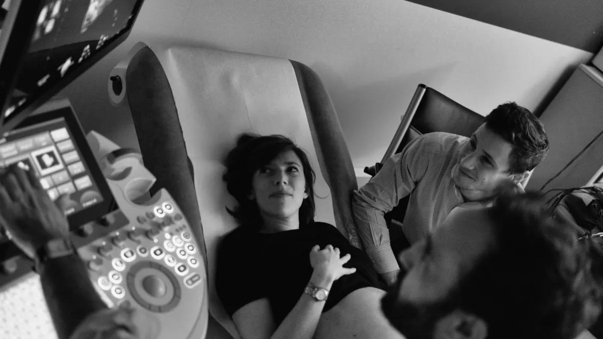
During the ultrasound, you will be lying down comfortably on your back on an examination table, with the lights turned down to facilitate viewing of the images displayed on the screen. Before beginning the exam, the sonographer will apply a gel to your abdomen which allows the sound waves to be transmitted. The ultrasound lets you see your baby for the first time and can be a very moving experience!
The Fetal Medicine Unit of the American Hospital of Paris has testing and diagnostic infrastructure featuring latest-generation ultrasound equipment and highly qualified sonographers who perform:
- Routine prenatal screening ultrasounds
- Specialized second-line diagnostic ultrasounds
These ultrasound exams are used to identify any fetal abnormalities. If an abnormality is detected, a prenatal consultation with a specialist will be proposed to determine if further genetic testing is necessary, such as chorionic villus sampling, amniocentesis or fetal blood sampling.
> Schedule an appointment with a sonographer or contact the Fetal Medicine Unit to make an appointment:
+33 1 46 41 28 82
centre-diagnostic-prenatal@ahparis.org